17 July 2016

Lecture #13: SUBPHYLUM SARCODINA


Species belonging to this group are the amoeba. This parasite move by simple extrusion of the protoplasm known as pseudopodia, which is also use for food collection.

General rules in identifying amoeba:

1.      All amoebas are non–pathogenic except E. histolytica.
2.      All amoebas are found in colon or large intestine except E. gingivalis.
3.      All amoebas can develop cyst and trophozoite except E. gingivalis & D. fragilis.

Based on nuclear characteristic, the Genus is divided into:

1.      Entamoeba – characterized by a small karyosome near the center of the nucleus and numerous chromatic granules lining the nuclear membrane.

a.      E. histolytica
b.      E. coli
c.       E. gingivalis  

2.      Endolimax – characterized by a relatively large karyosome of irregular shape and several achromatic threads connecting it with a delicate nuclear membrane.

a.      E. nana

3.      Iodamoeba – characterized by a large karyosome (endosome) rich in chromatin, typically surrounded by a single layer of periendosomal granules and attached to the karyosome and the nuclear membrane by radiating achromatic fibrils.

a.      I. butschlii

4.      Dientamoeba – characterized by karyosome consisting of several chromatin granules embedded in an achromatic matrix which is connected with a delicate nuclear membrane by a very fine achromatic fibrils.

a.      D. fragilis


******  ENTAMOEBA HISTOLYTICA  ******

Diseases:       Amoebiasis, amoebic dysentery, amoebic colitis

Morphology:

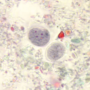
1.      Cyst – 3–20u, quadrinucleated, thin nuclear membrane, cigar–shaped chromatoidal body


2.      Trophozoite – 15–60u, long, fingerlike pseudopodia, progressive directional motility, nucleus with central karyosome, “Bull’s eye karyosome,”cytoplasm contains RBC.


Stool consistency and its association with stage of amoeba recovery

Two varieties:

1.      E. histolytica – larger, mean size is more than 10u; pathogenic

2.      E. hartmanii – small race, mean size is less than 10u; non–pathogenic

Comparison of cyst and trophozoite


                                                Trophozoite                          Cyst

Size
            Small race                  5 – 12u                                    3 – 10u
            Large race                  12 – 40u                                  10 – 16u

Motility & pseudopodia;    active, progressive &           spherical
shape of cyst             directional; finger–like,
                                                rapidly extruded

Unstained organism                  motile or rounded;              1–4 nuclei not visible;
in saline                     nucleus not visible              chromatoid matter refractile
                                    RBC visible                           glycogen refractile in young
                                                                                    cyst

Iodine stained                       nucleus visible                     nuclei visible; chromatoids
                                                                                                seldom seen; glycogen ball
                                                                                                in young cysts

hematoxyline stained          nucleus visible                     nuclei visible; chromatoids
                                                                                    stained; glycogen unstained

Inclusions                              RBC, in amoebic dysentery;                       None
                                                no bacteria when in fresh stool;
                                                no RBC in small race

Chromatoid matter              None                                      Rods with rounded end
                                                                                                (cigar–shaped); irregular
                                                                                                in small race

Glycogen                               None                                      diffuse in older cysts; mass
                                                                                                in cysts with 1–2 nuclei

Nucleus                                 single; ring–like                   1–4 may be present
                                                visible in stained
                                                preparations

Nuclear membrane              delicate; head–like               same as trophozoite
                                                chromatin dots

Karyosome                            small & centrally located    same as trophozoite

Life cycle


******  ENTAMOEBA GINGIVALIS  ******

Morphology:

1.      Only the trophozoite stage has been found and encystation probably does not occur.


2.      Has multiple pseudopodia; endoplasm contains phagocytosed and partly digested host leukocytes epithelial cells, at times bacteria and spirilla; spherical nucleus with a distinct membrane lined with a closely packed chromatin granules and near the center, a moderately well – defined karyosome, with delicate achromatic strands extending to the nuclear membrane.

Habitat

Gingival tissues around the teeth especially in pyorrhea alveolaris, but also thrives in apparently hygienic mouths or on dental plates if they are not kept meticulously clean.

Mode of reproduction       Binary fission


******  ENTAMOEBA COLI  ******
    

                                            Trophozoites                                                Cysts

Size                                         15–50u                                                10–30u

Motility & pseudopodia     sluggish, rarely progressive          spherical
            shape of cyst             & directional; blunt

Unstained organism            motile or rounded; nucleus           1–8 nuclei visible;
            in saline                     visible in endoplasm behind         chromatoid matter
                                                pseudopod; vacuoles seen             refractile; glycogen
                                                                                                            refractile in young cyst

iodine stained                       nucleus visible                                 nuclei visible;
                                                                                                            chromatoids seldom
                                                                                                            seen; glycogen ball

Hematoxyline stained         nucleus visible                                 nuclei visible;
                                                                                                            chromatoids stained
                                                                                                            glycogen unstained

Inclusions                              bacteria and food debris                 None
                                                no RBC

Chromatoid matter                                                                          bats with splinter ends;
                                                                                                            some comma or needle
                                                                                                            shaped
Glycogen                               None                                                  Diffuse in older cysts
                                                                                                            mass with 1–2 nuclei

Nucleus                                 single; ring–like; visible                 1–8 may be present
                                                in all preparations

Nuclear membrane              thick, coarse, irregular                    same as trophozoite
                                                chromatin dots

karyosome                            large eccentrically located              same as trophozoite


******  ENDOLIMAX NANA  ******


                                                Trophozoite                                      Cysts

Size                                         6–15u                                                  5–14u

Motility & pseudopodia     sluggish; hyaline budding             ovoidal or spherical
            shape of cyst

unstained organism in        motile or rounded; nucleus           1–4 nuclei not visible
            saline                          not visible; vacuoles seen

Iodine stained                       nucleus not visible                          nuclei visible

Hematoxylin                         nucleus visible; cytoplasm                         nuclei visible; no
                                                Vacuolated                                        chromatoids seen but
                                                                                                            bacilliform bodies
                                                                                                            maybe seen  

Inclusions                              bacteria & food debris                     none
                                                no RBC

Chromatoid matter              None                                                  None

Glycogen                               None                                                  diffuse; rarely has a
                                                                                                            mass in young cysts

Nucleus                                 single; visible with hematoxylin   1–4 maybe present
                                                and other permanent stain only

nuclear membrane               chromatin seldom present              same as trophozoite

karyosome                            large, central or eccentric                same as trophozoite


******  IODAMOEBA BUTSCHLII  ******


                                                Trophozoite                                      Cysts


Size                                         6–20u                                                  5–20u

Motility & pseudopodia     sluggish; hyaline; blunt                  irregular
            shape of cyst             slowly extruded      

Unstained organism in        motile or produced nucleus          nucleus not visible;
            saline                          not visible                                         glycogen refractile

Iodine stained                       nucleus not visible                          nucleus not visible

Hematoxylin stained           nucleus visible; cytoplasm                         nucleus visible; volutin
                                                            vacuolated                            granules stained;
                                                                                                            glycogen ball unstained

Inclusion                                bacteria & food debris;                    None
                                                no RBC

Chromatoid matter              None                                                  None

Glycogen                               None                                                  usually present as a
                                                                                                            well–defined mass

Nucleus                                 single; visible with hematoxylin   single
                                                & other permanent stains only

Nuclear membrane              same as E. nana                                same as trophozoite

Karyosome                            large, central or eccentric;               large; usually eccentric;
                                                surrounded by refractile,                achromatic granules on
                                                achromatic granules                        side of karyosome


******  DIENTAMOEBA FRAGILIS  ******


Disease:         Dientamoeba diarrhea or dientamebiasis

Morphology:


            Size                                                     :           5 – 12u
            Motility & pseudopodia                 :           active, hyaline, blunt & leaflike
            Unstained organism in saline        :           motile or rounded, nuclei not seen
            Iodine stained                                   :           stained not visible
            Hematoxylin stained                       :           nuclei seen
            Inclusion                                            :           bacteria & food debris; yeast; no RBC
            Chromatoid matter & glycogen     :           none
            Nucleus                                             :           Single but more frequently two
            Nuclear membrane                          :           delicate but no chromatin dot seen
            Karyosome                                        :           fragmented usually into 4 granules
                                                                                    or tetrakaryosome or fragmented

                                                                                    karyosome 


No comments: