
The hookworms are characterized by the presence of cutting plates
which may either be teeth or semilunar notch. This characteristic
differentiates them from other nematodes.

************ Human hookworms ************

1.
Necator americanus (New World Hookworm) – located in the jejenum
a.
Mode of transmission: skin penetration
b.
Infective stage: Filariform larva
c.
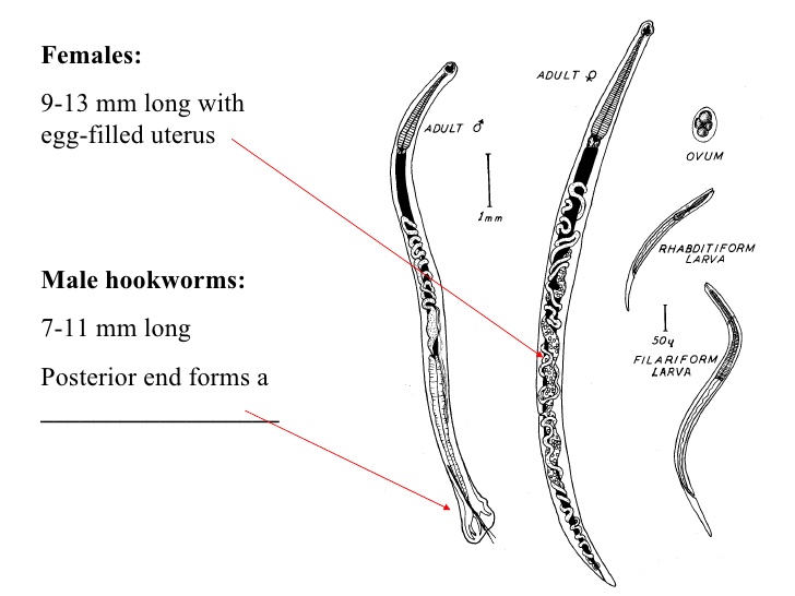
Adult worms are cylindrical in shape with
cephalic portion curve against the curve of the body and have a small buccal
capsule with a pair of semilunar cutting plates.
2.
Ancylostoma duodonale (Old World Hookworm) –
located in the duodenum
a.
Adult worm is stout, cylindrical, slightly
constricted anteriorly and has a cephalic curvature along the curve of the
body. Living worms are pinkish or creamy–gray in color and are covered with a
tough cuticula and are provided with a pair of small teeth (accessory) and 2
pairs of big teeth.

N.B: The life cycle, disease produced, diagnosis, treatment and
ova
morphology
of Necator americanus and Ancylostoma sp. are the same,
they
only differ in the morphology of adult male and female.
Morphology of the
ova: (1) Ovoid (2) Transparent (3) with 2–8 germ cells

************ Animal hookworms ***********
1.
Ancylostoma braziliense (cat hookworm)
a.
Disease produced: Cutaneous larva migrans
(creeping eruption)
b.
The buccal capsule is specifically
diagnostics, having a pair of small, very inconspicuous median teeth and a pair
of larger outer teeth.
2.
Ancylostoma caninum (dog hookworm)
a.
Disease produced: Cutaneous larva migrans
(creeping eruption)

b.
The adult has a wide buccal capsule to accommodate
3 pairs of ventral teeth, a diagnostic characteristic of the species.

3.
Gnathostoma spinigerum
a.
Gnathostomiasis is caused by infection with
the larval stage of the parasite. Adult stage is normally found in the tumor of
the stomach of wild and domesticated cats and dogs.


b.
The adult is curved ventrally on both ends,
stout, reddish in color and slightly transparent with a subglobuse cephalic
swelling with 4–8 rows of spines transversely arranged, with a neck or cephalic
constriction and with spines from cuticle in anterior half of the body.
c.
Mode of transmission is by skin penetration
4.
Angiostrongylus cantonensis (rodent lungworm)
a.
Causes eosinophilic meningitis, cerebral
angiostrongyliasis

b.
Definite host: Rattus rattus, R. norvegious
c.
Snail host: Achatina fulica (giant African snail),
Pila polita, P. gracius, P. ecpansa
No comments:
Post a Comment