
A cell is the basic structural, functional, and biological unit of
all known living organisms. A cell is the smallest unit of life that can
replicate independently, and cells are often called the “building blocks of
life.” When a cell is exposed to an injurious agent, the possible results are:
a.
The cell may adapt to the situation or
b.
The cell may acquire a reversible injury
c.
The cell may obtain an irreversible injury
and may die. The cell may die via one of two ways: either by necrosis or by
apoptosis.
Types of cellular adaptation

A.
Hypertrophy
Hypertrophy is increase in the
size of cells. Increase workload leads to increased protein synthesis and
increased size and number of intracellular organelles which, in turn, leads to
increased cell size. The increased cell size leads to increased size of the
organ.
Example: cardiomegaly
B.
Hyperplasia
Hyperplasia is an increase in the
number of cells. It can lead to an increase in size of the organ. It is usually
caused by hormonal stimulation. It can be physiological as in enlargement of
the breast during pregnancy or it can be pathological as in endometrial
hyperplasia.
C.
Atrophy
Atrophy is a decrease in the size
of the cell. This can lead to decreased size of the organ. The atrophic cell
shows autophagic vacuoles which contain cellular debris from degraded
organelles.
Atrophy can be caused by: (1) Disuse
(2) Undernutrition (3) Decreased endocrine stimulation (4) Denervation (5) Old
age
D.
Metaplasia
Metaplasia is the reversible
replacement of one differentiated cell type with another mature differentiated
cell type. The change from one type of cell to another may generally be a part
of normal maturation process or caused by some sort of abnormal stimulus.
In simplistic terms, it is as if
the original cells are not robust enough to withstand the new environment and
so they change into another type more suited to the new environment. If the
stimulus that caused metaplasia is removed or ceases, tissues return to their
normal pattern of differentiation.
Types of Cell Damage
A.
Fatty Change
Cell has been damaged and is
unable to adequately metabolize fat. Small vacuoles of fat accumulate and
become dispersed within cytoplasm. Mild fatty change may have no effect on cell
function; however more severe fatty change can impair cellular function. In the
liver, the enlargement of hepatocytes due to fatty change may compress adjacent
bile canaliculi leading to cholestasis. Depending on the cause and severity of
the lipid accumulation, fatty change is generally reversible.
B.
Cellular Swelling
Cellular swelling may occur due
to cellular hypoxia, which damages the sodium–potassium membrane pump; it is
reversible when the cause is eliminated. Cellular swelling is the first
manifestation of almost all forms of injury to cells. When it affects many
cells in an organ, it causes some pallor, increased turgor, and increase in weight
of the organ. On microscopic examination, small clear vacuoles may be seen
within the cytoplasm; these represent distended and pinched – off segments of
the endoplasmic reticulum. This pattern of non – lethal injury is sometimes
called hydropic change or vacuolar degeneration. The ultrastructural changes of
reversible cell injury include: (1) Blebbing (2) Blunting (3) Distortion of
microvilli (4) Loosening of intercellular attachments (5) Mitochondrial changes
(6) Dilatation of the endoplasmic reticulum.
C.
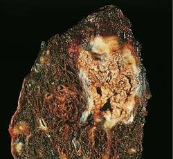
Necrosis
Necrosis is characterized by
cytoplasmic swelling, irreversible damage to the plasma membrane and organelle
breakdown leading to cell death. Necrosis does not occur in dead organisms. In
dead organisms, autolysis and heterolysis take place.
Stages of Cellular Necrosis:
a.
Pyknosis – is the irreversible condensation
of chromatin in the nucleus of a cell undergoing necrosis or apoptosis.
b.
Karyorrhexis – is the destructive
fragmentation of the nucleus of a dying cell.
c.
Karyolysis – is the complete dissolution of
the chromatin of a dying cell due to the enzymatic degradation by
endonucleases.
Types of Necrosis:
a.
Coagulative Necrosis – is a result of sudden
interruption of blood supply to an organ especially to the heart causing
ischemia or infarction. It can also be induced by high local temperature such
as the effect of high intensity focused ultrasound as applied to cancerous
cells.

b.
Liquefactive Necrosis – is a result of a
transformation of the tissue into a liquid viscous mass. Often it is associated
with focal bacterial or fungal infections. In liquefactive necrosis, the affected
cell is completely digested by hydrolytic enzymes, resulting in a soft,
circumscribed lesion consisting of pus and the fluid remains of necrotic
tissue. Dead leukocytes will remain as creamy yellow pus. After the removal of
cell debris by white blood cells, a fluid filled pace is left. It is generally
associated with abscess formation and is commonly found in the central nervous
system.

c.
Fat Necrosis – the enzyme lipase releases
fatty acids from triglycerides. The fatty acids then complex with calcium to
form soaps. These soaps appear as white chalky deposits. It is usually
associated with trauma of the pancreas or acute pancreatitis. It can also occur
in the breast, the salivary glands and neonates after a traumatic delivery.

d.
Caseous Necrosis – is a form of cell death in
which the tissue maintains a cheese–like appearance. The dead tissue appears as
a soft and white proteinaceous dead cell mass. Frequently, it is encountered in
the foci of tuberculosis infection. It can also be caused by syphilis and
certain fungi.
e.
Gangrenous Necrosis – is a type of necrosis caused
by a critically insufficient blood supply. This potentially life–threatening
condition may occur after an injury or infection, or in people suffering from
any chronic health problem affecting blood circulation. The primary cause of
gangrene is reduced blood supply to the affected tissues, which results in cell
death. Diabetes and long–term smoking increase the risk of suffering from
gangrene.
(1) Dry Gangrene is a form of coagulative necrosis
that develops in ischemic tissue, where the blood supply is inadequate to keep
tissue viable.

(2) Wet Gangrene is characterized by thriving
bacteria and has a poor prognosis (compared to dry gangrene) due to sepsis
resulting from the free communication between infected fluid and circulatory
fluid. In Wet Gangrene, the tissue is infected by saprogenic microorganisms
(Clostridium perfringens and Bacillus fusiformis) which cause tissue to swell
and emit a fetid smell. The affected part is saturated with stagnant blood,
which promotes the rapid growth of bacteria. The toxic products formed by
bacteria are absorbed, causing systemic manifestation of sepsis and finally
death. The affected part is edematous, soft, putrid, rotten and dark.
(3) Gas Gangrene is a bacterial infection that
produces gas within tissues. It is mostly caused by alpha toxin producing
Clostridium perfringens. Infection spreads rapidly as the gases produced by
bacteria expand and infiltrate healthy tissue in the vicinity. Because of its
ability to quickly spread to surrounding tissues, gas gangrene should be
treated as a medical emergency. Gas gangrene can cause necrosis, gas production
and sepsis. Progression to toxemia and shock is often very rapid.
(4) Necrotizing
Fascitis is an
infection of the deeper layer of skin and subcutaneous tissues caused by
organisms that normally reside on the individual’s skin. They destroy the
tissue that makes up the skin and muscle by releasing toxins (virulence factor)
which include streptococcal pyogenic exotoxins.

D.
Apoptosis
Apoptosis is a process of
programmed cell death that occurs in multicellular organisms. Apoptosis usually
occurs as a physiologic process for removal of cells during embryogenesis,
menstruation, etc.
Excessive apoptosis causes
atrophy, whereas an insufficient amount results in uncontrolled cell
proliferation, such as cancer. Some factors like Fas receptors and caspases
promote apoptosis while some members of the Bcl–2 family of proteins inhibit
apoptosis.

Pathologic Calcification
Calcification of soft tissue (arteries, cartilage, heart valves,
etc.) can be caused by Vitamin K2 deficiency or by poor calcium absorption due
to high Calcium / Vitamin D ratio. This can occur with or without a mineral imbalance.
Intake of excessive Vitamin D can cause Vitamin D poisoning and
excessive intake of Calcium from the intestine, when accompanied by a
deficiency of Vitamin K (perhaps induced by an anticoagulant) can result in
calcification of arteries and other soft tissue. Such metastatic soft tissue
calcification is mainly in tissues containing “calcium catchers” such as
elastic fibres or sour mucopolysaccharides. These tissues especially include
the lungs (pumice lung) and the aorta.
A.
Dystropic Calcification is caused by abnormalities or
degeneration of tissues resulting in mineral deposition, though blood levels of
calcium remain normal. These differences in pathology also mean that metastatic
calcification is often found in many tissues throughout a person or animal,
whereas dystrophic calcification is localized.
B.
Metastatic Calcification is deposition of calcium salts
in otherwise normal tissue, because of elevated serum levels of calcium, which
can occur because of deranged metabolism as well as increased absorption or
decreased excretion of calcium and related minerals, as seen in
hyperthyroidism.


No comments:
Post a Comment